
✅Medically reviewed
Erections depend on blood. Healthy arteries open wide, and healthy veins close tight. When either system fails, no tablet or injection can replace an accurate diagnosis.
Many men reach a private clinic after months of guesswork. Some try tablets from online pharmacies. Others read about regenerative options such as the P shot. Fewer men ask whether a scan should come first.
A penile doppler ultrasound answers that question with measurements rather than assumptions. The scan tracks blood moving into the penis and leaving it again. It shows whether the problem sits in the arteries, the veins, or neither.
That detail matters for any man considering P shot treatment. The P shot uses platelet-rich plasma (PRP) drawn from the patient’s own blood. Clinical evidence remains limited. Careful selection therefore carries real weight.
This article explains what the scan measures and when guidelines recommend it. Further sections examine the evidence for PRP and the limits of imaging. Sources include the NHS, NICE and peer-reviewed journals. Readers finish with a clear framework for an informed decision.
What Is a Penile Doppler Ultrasound?
This imaging test measures blood flow in the penile arteries using sound waves. Specialists also call it penile duplex Doppler or colour Doppler ultrasonography. Two techniques work together. Grey-scale imaging shows structure. Doppler imaging shows the speed and direction of moving blood.
A 2019 review in the American Journal of Roentgenology covers technique and interpretation in detail. The scan does more than photograph anatomy; it measures how the penis behaves during an erection.
Why Blood Flow Matters
An erection follows a precise vascular sequence. Nerve signals relax smooth muscle in the cavernosal arteries. Blood fills the erectile chambers and compresses the draining veins. Pressure rises, and rigidity follows. Any disruption in inflow or venous trapping weakens the result. Doppler imaging measures exactly these two steps.
How the Scan Works
The test follows a fixed sequence:
- A clinician applies gel and places a high-frequency probe on the penis.
- Baseline images record flaccid anatomy and blood flow.
- A vasoactive drug then induces an erection. Alprostadil, papaverine or a combination are common choices.
- Repeated measurements follow at set intervals until the erection peaks.
- Staff monitor the patient until the erection subsides.
Specialist clinics sometimes repeat the drug dose when the response looks incomplete.
What the Numbers Mean
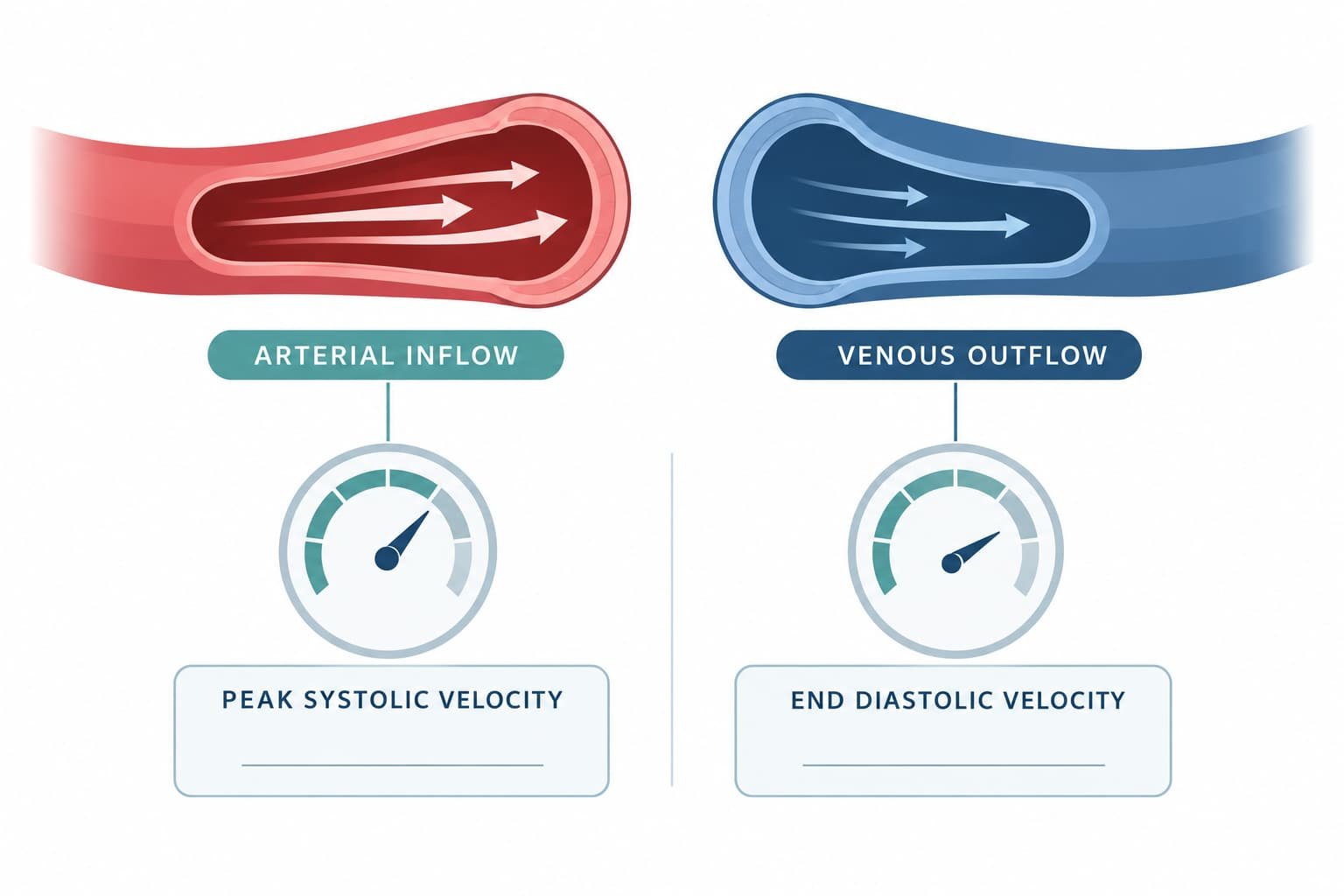
Two measurements carry most of the weight. Peak systolic velocity (PSV) reflects arterial inflow. End diastolic velocity (EDV) reflects venous leakage. Urological protocols commonly treat a PSV above 30 cm/s as normal. An EDV below 5 cm/s suggests an intact veno-occlusive mechanism.
Low PSV points towards arterial insufficiency. High EDV points towards venous leak. Many reports also include the resistance index as a cross-check.
Flores, West and Mulhall published a protocol in the Journal of Sexual Medicine in 2024. Accuracy depends on complete relaxation of the cavernosal smooth muscle. A redosing protocol improves reliability.
Who Performs the Scan and What the Report Contains
Consultant radiologists and urologists with andrological training usually perform the scan. Training matters because technique shapes every measurement. A complete report lists several items:
- Dose and type of vasoactive agent used
- Peak systolic velocity and end diastolic velocity in both cavernosal arteries
- Degree of rigidity achieved at each time point
- Any structural abnormality, such as plaque or fibrosis
- Final interpretation with clinical correlation
Men can request a copy of the report. Referring clinicians use that document to plan management. A report without dose or rigidity details limits interpretation.
Erectile Dysfunction Diagnosis: Where Does the Scan Fit?
Most erectile dysfunction diagnosis starts in primary care. NICE Clinical Knowledge Summaries advise a sexual history, a medication review and a physical examination. Blood pressure, cardiovascular risk and blood tests complete the first assessment.
The NHS lists diabetes, high blood pressure and narrowed arteries among common causes. Erectile dysfunction can act as an early marker of vascular disease. Researchers link this to the small calibre of penile arteries.
Erectile Dysfunction Tests UK
Erectile dysfunction tests UK patients encounter fall into clear tiers:
- Questionnaires such as the IIEF-5 score symptom severity.
- Laboratory tests check glucose or HbA1c, cholesterol and morning testosterone.
- Blood pressure and weight readings estimate vascular risk.
- Specialist assessment adds Doppler imaging, nocturnal tumescence testing or cavernosometry.
Most men need only the first three assessments. Referral for specialist testing follows when first-line treatment fails or the history raises concern.
Doppler Ultrasound for Erectile Dysfunction: When Do Guidelines Recommend It?
Penile Doppler ultrasound for erectile dysfunction is not a routine first test. Specialist guidance, including European Association of Urology recommendations, limits vascular imaging to selected men. Typical indications include:
- Lifelong erectile dysfunction in a young man
- A history of pelvic or perineal trauma
- Suspected Peyronie’s disease or penile curvature
- Assessment before vascular or penile surgery
- Poor response to first-line PDE5 inhibitors such as sildenafil
Vascular risk factors add further weight. Diabetes, hypertension and high cholesterol all raise the probability of a vascular cause. Reviews describe the scan as especially useful when oral medicines fail.
What a Scan Can Diagnose
- Arterial insufficiency from narrowed or stiff cavernosal arteries
- Venous leak, also called veno-occlusive dysfunction
- Normal vascular findings, which point towards psychological or non-vascular causes
- Structural changes such as plaques, fibrosis or calcification
Published figures show the range of results. A Polish Journal of Radiology study examined 88 men with erectile dysfunction. Abnormal Doppler findings appeared in 57 per cent. Venous insufficiency accounted for most abnormal results.
Does a P Shot Candidate Need a Scan First?

No NHS or NICE guidance makes the scan compulsory before a P shot. The decision depends on history, examination and the clinical question. A penile doppler ultrasound helps most when the cause of erectile dysfunction remains unclear.
How Clinicians Combine Findings
Clinicians rarely rely on one result. A diagnostic picture combines symptoms, risk factors, examination findings and test data. Consistent findings across sources raise confidence. Conflicting findings prompt further assessment. Sudden onset with normal morning erections, for example, points more towards a psychological cause. Imaging adds little in that scenario.
Why a Scan Can Help Before P Shot Treatment
Proponents suggest that PRP releases growth factors that may support tissue repair and new vessels. This theory remains under study. Objective vascular data therefore add useful context.
- Baseline data. Doppler measurements record blood flow before treatment and allow objective comparison afterwards.
- Cause identification. Severe arterial disease or venous leak may limit any response, so expectations need adjusting.
- Structural findings. Imaging detects Peyronie’s plaques or calcification that need separate management.
- Cardiovascular safety. Abnormal arterial findings prompt a wider cardiovascular review.
Why a Scan May Not Be Necessary
Many men present with a clear, mild picture. Gradual onset, partial erections and known risk factors make a vascular cause likely. In such cases, history, examination and blood tests may suffice.
The scan also carries burdens. It requires a drug-induced erection and a private clinical setting. Risks include pain, bruising, dizziness and, rarely, priapism. Cost and waiting times add further limits. Clinicians therefore weigh benefit against burden for each patient.
When to Seek Urgent Help
An erection lasting more than four hours needs emergency care. The NHS advises attending A&E immediately in that situation. Delay risks permanent tissue damage.
Limitations of the Scan
- Clinic anxiety can blunt the drug response and produce false arterial readings.
- Incomplete smooth muscle relaxation distorts measurements.
- Operator skill strongly affects technique and interpretation.
- No validated Doppler threshold predicts response to PRP.
A 2020 Journal of Sexual Medicine paper reviewed methodological pitfalls and interpretation deficiencies in duplex Doppler studies. Scan results guide clinical judgement but never replace it.
Other Specialist Tests Compared
Doppler imaging is not the only specialist option. Nocturnal penile tumescence testing records erections during sleep and helps separate psychological from organic causes. Cavernosometry measures pressure and flow directly through needles placed in the penis. This test is invasive, so clinicians reserve it for selected cases. Questionnaires and blood tests remain the foundation of every assessment.
Special Groups Worth Noting
Certain groups need particular attention. Men with diabetes often show reduced arterial inflow alongside nerve damage. A scan documents the vascular component but cannot measure neuropathy. Men with Peyronie’s disease may have plaques that ultrasound can map. Radical prostate surgery often causes erectile dysfunction through nerve injury, so Doppler findings may look normal.
Each group needs an individual plan. Evidence for PRP in these groups remains limited. A clinician explains which findings matter most for each situation.
What Evidence Supports the P Shot?
The P shot involves injecting PRP into the penis. Evidence remains early. Small randomised trials report improved erectile function scores after PRP. A 2021 double-blind, placebo-controlled trial by Poulios and colleagues is one example. Sample sizes stay small and follow-up stays short. Researchers also debate PRP preparation methods, platelet concentration and injection schedules. This variation complicates comparison between studies.
Specialist guidelines currently regard PRP for erectile dysfunction as investigational. NICE has not issued guidance recommending it. The NHS does not routinely fund it. Claims about P shot UK outcomes deserve careful scrutiny.
Reported adverse events remain mild. Examples include bruising, swelling and transient pain at injection sites. Because PRP derives from the patient’s own blood, allergic reactions appear unlikely. Infection remains a rare risk with any injection. Long-term safety data remain limited.
Private providers deliver P shot treatment within a regulated framework. The Care Quality Commission registers independent clinics in England. Separately, the General Medical Council keeps the register of licensed doctors. Men can verify both online.
Choosing a Clinic for P Shot London Assessment
P shot London providers cluster around Harley Street and Marylebone. Dr Syed Nadeem Abbas leads Pshots clinic UK in Marylebone. Qualifications comprise MBBS, MRCS RCS Edinburgh and MRCGP. An MSc in Aesthetic Plastic Surgery with Distinction came from Queen Mary University of London. Training took place at Cambridge, Oxford and the Royal London Hospital.
Men can ask any provider whether specialist imaging suits the individual situation. Practitioners should explain indications, risks and uncertainty before any treatment decision.
Questions to Raise at a Consultation
Structured questions improve decisions. Useful examples include:
- Which tests confirm the cause of erectile dysfunction?
- Does the clinical history justify a scan?
- Would any finding exclude P shot treatment?
- How will outcome measures track response?
- What qualifications does the professional who performs the scan hold?
- Where should urgent symptoms go after any procedure?
Written answers help men compare providers fairly. Clear, consistent responses signal careful practice.
Preparing for the Scan Appointment
Before the Appointment
Clinics give written instructions. Men disclose all medicines, especially anticoagulants, along with allergies and heart conditions. A warm, private room reduces anxiety and supports a reliable result.
During and After the Scan
Appointments commonly last 30 to 60 minutes. Staff stay until the erection fully subsides. Mild discomfort from the injection is typical. Bruising may appear at the injection site.
Realistic Outcomes and Alternatives
A normal scan does not guarantee P shot success. An abnormal scan does not exclude it. Individual responses vary, and long-term data remain limited. Some protocols propose repeated sessions, which adds cost.
Follow-up measurement completes the picture. Repeat questionnaires such as the IIEF-5 document change over time. Follow-up imaging remains optional and depends on clinical need. Objective tracking protects against over-interpretation of short-term improvement.
Other options carry stronger evidence. The NHS lists PDE5 inhibitors such as sildenafil as first-line medicines. Lifestyle change, psychosexual counselling, vacuum devices and injections also feature in standard pathways. Cardiovascular risk control improves general health regardless of erection outcome.
Cost and access deserve mention. Private scans carry separate fees that vary between providers. Waiting times for NHS imaging vary by region and urgency. Men weighing a P shot London pathway can request an itemised estimate covering assessment, imaging and any repeat sessions. Transparent pricing supports fair comparison.
Common Misunderstandings
- A scan treats erectile dysfunction. In fact, the scan only diagnoses.
- A normal result proves the problem is psychological. Nerve, hormonal and drug-related causes still exist.
- An abnormal result rules out every treatment. Many options remain available.
- Every P shot candidate needs a scan. Guidelines reserve imaging for selected men.
Frequently Asked Questions
Is a penile doppler ultrasound painful?
Discomfort usually comes from the injection that induces the erection. It is typically mild and brief. The probe itself causes little discomfort.
Does the NHS offer Doppler ultrasound for erectile dysfunction?
NHS urology and radiology services offer it to selected men after referral. Availability varies between trusts. A GP carries out the first assessment.
Can the scan predict P shot success?
Current evidence says no. Researchers have not validated any Doppler threshold for predicting PRP response. The scan clarifies the underlying cause instead.
Is the P shot available on the NHS?
The NHS does not routinely fund PRP for erectile dysfunction. Private clinics provide it. Costs vary between providers.
Who should avoid the scan?
Clinicians take extra care with bleeding disorders, sickle cell disease and prior priapism. Allergy to the injected agents also matters. A doctor decides after reviewing the full medical history.
How long does a penile doppler ultrasound take?
Most appointments last between 30 and 60 minutes. The exact length depends on how quickly the erection develops.
Key Takeaways
Diagnosis should precede treatment. A penile doppler ultrasound offers objective data on arterial inflow and venous leakage. The scan suits selected men rather than every P shot candidate.
Good decisions combine history, examination, blood tests and, when justified, imaging. Evidence for PRP remains early. Realistic expectations protect men from disappointment and unnecessary cost.
Anyone considering P shot treatment benefits from a documented diagnosis. Written records support follow-up, second opinions and honest assessment of progress.
Verified credentials, transparent explanations and balanced evidence mark trustworthy care. Men who ask direct questions make better choices.
When blood flow drives the problem, should measurement not come before any injection?
Read more:
Tests to Take Before a P Shot: HbA1c, Testosterone, Lipids and Blood Pressure
Priapus Shot London: A Complete Guide for Patients Considering Treatment
Dr Syed Nadeem Abbas, MBBS, MRCSEd, MSc Aesthetic Plastic Surgery (Distinction) Medical Director, Pshot Clinic | CQC Regulated | Monday to Saturday 10:00–18:00 +44 7955 836986


